So let’s reflect on The Cell!
Hey guys, Richie’s here! Let’s get the ball rolling!
Lectures, tutorials all greeted by hungry minds starved by the Christmas vacation,, proved to be nothing short of stimulating. It was a breath of relief when I learnt that the pass rate last year for this course was 90%! But then again, I am not aiming just to pass this course. 🙂 Blogging was a word I met browsing the internet, reading books, but never did I believe that I would have been given this task for a lovely percentage of my Biochemistry course! The fear of the unknown was my initial reaction. Now as I am reflecting on my progress thus far, I am grateful for this opportunity (and the fact that it is a group assignment!). It gives us, the Biochemistry3RST team, the perfect avenue to display our academic and creative skills for the entire global audience! (You can probably tell how excited I am based on the amount of exclamation marks I used hitherto.) Each week you can look forward to pieces from fresh, alternating minds and this continuous renewal of our human resource is what will keep this blog inundating your minds with pretty cool Biochem stuff. 🙂

The first science I learnt in high school was the cell. My first note began something like this: “The cell was discovered by an English inventor and scientist called Robert Hooke who described the cork from the bark of a tree as being made up of thousands of tiny boxes. He called these boxes cells.” It’s still embedded in my memory after about seven odd years of teenage woes, ups and overcoming academic barriers. I remember seeing the name Robert Hooke on the Wednesday of our second lecture and I became overwhelmed by memories of learning the descriptions of these fascinating cellular organelles that build my interest which led me onto this path to a Biochemistry major.

Robert Hooke, the first person to use the word “Cell”.
This blog is to foster a love for a subject which has a rep for being tough, and the producer of low GPA’s. Upon saying so, it’s time for me to get down to earth with some of the greatly underrated yet awesome organelles! We all heard about the nucleus. It is perhaps the first sub-cellular component that we all knew existed, even without formal knowledge of life science, it being the largest organelle and the cell’s brain… Also the mitochondrion being the intracellular power station as it synthesizes ATP the energy currency and its characteristic geographical structure… (Stalactites and stalagmites!) So what about the ribosomes and other protein processing organelles? These tiny yet essential structures (ribosomes) synthesize proteins which are the bases for enzyme activity, your hair and nails, and other intriguing aspects of life, I mean, even your DNA are nucleic proteins!

DNA wrapped around histones forming nucleosomes… How interesting!!

The might mitochondrion!! lol
This organelle that has to be seen with an electron microscope consist of Ribosomal RNA (rRNA) and is made in the nucleolus (within the nucleus). A ribosome has a large and small subunit and it is the latter that receives messenger RNA (mRNA) that is transcribed from your DNA in your nucleus. This mRNA feeds data about how to make proteins into the ribosomes which assemble them from amino acids.

Assembly of proteins in a ribosome with mRNA in action!
In eukaryotic cells (cells that contain membrane bound organelles and are between 10-100 µm) ribosomes can be found concentrated on a single membrane organelle called the rough endoplasmic reticulum (the last part of the name was quite funny to me when I was 12!). The RER is connected to the outer membrane of the nucleus and synthesized proteins are collected in the spaces between its membranes called the cisternae. Here, these spruce proteins are then modified into glycoproteins and lipoproteins by the attachment of other biomolecules.

The ER connected to the nucleus’ outer membrane and studded with tiny ribosomes.
If a mutated gene is transcribed into mRNA, then the resulting protein assembled in RER will be an inaccurate, unacceptable variation of the protein. The implications of this is of great medical proportions as the protein will not be able to effectively carry out its purpose, or fail to do it entirely. Cystic fibrosis (CF) occurs when a particular cell membrane chloride channel protein (Cystic Fibrosis Transmembrane Regulator- CFTR) is not properly manufactured and is trapped in the Endoplasmic Reticulum and consequently degenerated. CF leads to an accumulation of mucus in bodily organs such as the pancreas and lungs. Furthermore, ‘stress’ of the Endoplasmic Reticulum due to accumulation of amino acids and fatty acids, lack of glucose and dwindling calcium ion supplies can ultimately trigger the cell’s destruction and it plays a role in infamous diseases such as Parkinson’s and Alzheimer’s.

Were you expecting a GORY image?! 😛
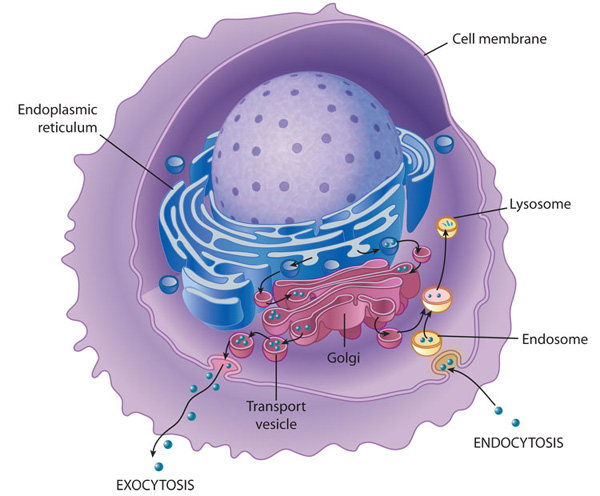
Proteins continue their intracellular journey when they ‘pinch of’ from the RER as a membrane bounded vesicle which embeds itself into the cis/convex face of the Golgi apparatus. Like a postal delivery service, the proteins and modified biomolecules are given an address and depending on the type of protein/enzymes they are, they can remain in the cell (after leaving the Golgi apparatus) as a lysosome which destroys pathogens or it may leave the cell entirely. Lysosomes may also break down proteins in the form of dying organelles. They accomplish these combats by fusing their single membrane with that of a vesicle containing the pathogen or retired organelle, consequently secreting their enzymes (proteases and lipases) which does the exterminating. Proteasomes sole function is to degrade intracellular proteins; they have no business with the proteins in your digestive system that you ate. These help out the lysosomes in recycling amino acids.

Now… Linking all the protein synthesizing and processing organelles
Medical horrors such as the Tay-Sachs disease and Pompe’s disease are lipid storage maladies caused by the absence of one highly essential lysosomal enzyme. Both of the diseases are unfortunately hereditary but in the case of the latter, this rear ailment affects the heart and skeletal muscles. A mutated gene codes for an alternate, dissimilar form of the enzyme- acid, alpha-glucosidase (GAA) that usually breaks down glycogen to glucose and is found in lysosomes. The absence of GAA would starve muscles for glucose and lead to an accumulation of lysosomal glycogen throughout the body especially in the heart and skeletal muscles. In both cases despite treatment death can occur at early childhood. 😦 Such a sad thing to write before concluding my first post on this blog…

Symptoms of this monster disease in tiny kids 😦
In retrospect, the chemistry of sub-cellular components being highly interesting is also quite relevant to modern science and medicine. Even though I focused my attention on the protein related organelles and diseases, the cell is a vast universe waiting for you to explore 🙂 and at this very moment research is being conducted in institutes worldwide research that can save lives as far as organelles and human diseases are concerned…. So my fellow Biocheminions, read, Read, READ, explore, Explore, EXPLORE to become the next generation of scientific heroes!!!

The cell is a vast universe waiting for you to explore!!!
Until I blog again, ACCENTUATE THE POSITIVE,
Richie, over and out!
References:
http://www.ninds.nih.gov/disorders/pompe/pompe.htm
http://www.mhhe.com/biosci/ap/foxhumphys/student/olc/h-reading10.html
D.J Taylor, N.P.O Green, G.W. Stout, 1984 Biological Science 1. Cambridge University Press.
Images and Animation references :
http://bi0l0gy.wikispaces.com/file/view/rough_endoplasmic_reticulum.jpg/275154994/rough_endoplasmic_reticulum.jpg- Rough Endoplasmic Reticulum
http://en.wikipedia.org/wiki/File:Protein_translation.gif – Ribosome animation
http://www.cancer.gov/PublishedContent/Images/images/targetedtherapies/mm/clip_image006_0002.jpg – Histones wrapped in histones
http://media-cache-ak0.pinimg.com/236x/f8/30/57/f830573809212927254614f2277a779e.jpg – Robert Hooke
http://www.nature.com/scitable/content/ne0000/ne0000/ne0000/ne0000/14713129/U3CP3-3_MembraneTransport_k.jpg – Linkage of Organelles
http://www.pompe.com/~/media/Pompe/Images/Unused/pc_symptoms_baby_hcp_03.jpg- Pompe’s Disease
http://dishingitdaily.files.wordpress.com/2013/02/brain-cell-the-universe-birth-of-a-cell-death-of-a-star-eye-nebula.jpg – Cell and the Universe
